研究主題:超解析技術之應用(Super-resolution architecture)-細胞中心粒中DAPs的結構模型
撰寫:學生陳冠彬
了解細胞分裂的過程對於癌症的研究是非常重要的一環,然而細胞分裂是一個非常複雜的過程。在不同的階段,都有不同的蛋白質牽涉其中,最終才能完成細胞的分裂。在動物細胞分裂的週期到G2 期(後期)與 M 期(終期)之間,也就是細胞進入有絲分裂的轉折點時,DAPs(中心粒遠端附屬物,Centriole distal appendages)的運作扮演了重要的角色。在DAPs中的不同蛋白質,分別有著不同的功能,而楊東霖老師的團隊致力於利用超解析顯微鏡(Super-resolution microscopy)來觀測DAPs的工作過程以及建立3D模型。
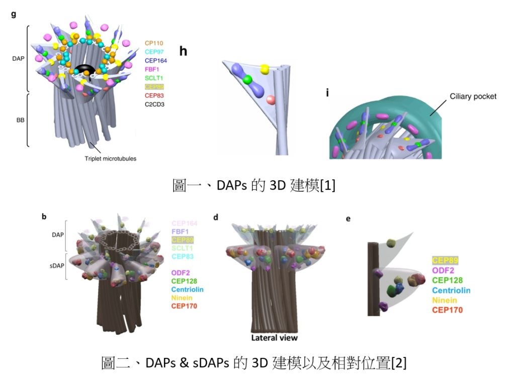
楊老師於2018年發表在自然通訊(Nature Communications)上的文章,就是利用超解析顯微鏡重建DAPs的3D模型。透過觀察正在 G0/G1階段的動物細胞,來定位每一個蛋白和它的功能。而動物細胞在此階段時,中心粒末端會生長出初級纖毛來提供多項細胞感測與訊號的傳遞。而DAPs在這個過程當中扮演了纖毛生成以及定位的工作。楊老師的團隊在這當中辨識出數個DAPs的功能蛋白質以及扇葉狀的9重對稱結構,成功利用單分子定位影像技術還原了DAPs的3D原貌,如圖一所示。
sDAPs(中心粒次遠端附屬物, subdistal appendages)是一個靠近DAPs的另外一個中心粒元素。楊老師於2020年在eLife期刊中提出了以往未曾發現關於sDAPs的生物機制。以往只發現了sDAPs可以在初級纖毛生長(ciliogenesis)時,提供紡錘絲錨定在微管(microtubule)上的支撐。而楊老師利用超解析顯微鏡發現DAPs跟sDAPs會產生耦合的現象。DAPs中的CEP89蛋白以及sDAPs中的ODF2蛋白存在著雙層的結構,會透過DAPs跟sDAPs的完整性來做調節,最終會影響DAPs跟sDAPs在中心粒遠端上的覆蓋區域,如圖二所示。
當我們能夠還原的生物機制越完整,在醫學上也可以更了解發病的原因,利用超解析顯微鏡觀察細胞的運作,不僅是更了解人體運作的原理,也是為了未來醫學鋪展更康莊的大道。
Understanding the process of cell division is an important part of cancer research; however, cell division is a very complex process. Different proteins are involved in various stages, ultimately leading to the completion of cell division. The operation of Centriole Distal Appendages (DAPs) plays an important role when animal cells enter the turning point of mitosis during the cell division cycle, between G2 phase (late phase) and M phase (end phase). Different proteins in DAPs have other functions, and Dr. Yang’s team is developing super-resolution microscopy to observe the working process of DAPs and establish 3D models.
Dr. Yang’s work in Nature Communications (2018) reconstructed the 3D model of DAPs using super-resolution microscopy. By observing mammalian cells undergoing mitosis, the location and function of each protein were determined. When mammalian cells undergo mitosis, the centriole ends in the centrosome and forms spindle fibers to provide the necessary pulling force for separating cells. DAPs play a role in generating and positioning cilia during this process. Dr. Yang’s team identified several functional proteins in DAPs and the 9-fold symmetric structure of the DAPs, successfully restoring the configuration of DAPs via 3D modeling, as shown in Figure 1.
Subdistal appendages (sDAPs) are another centriole element near DAPs. Dr. Yang proposed a previously unknown biological mechanism of sDAPs in eLife in 2020. Previously, it was only found that sDAPs may be associated with anchoring spindle fibers on microtubules during ciliogenesis. However, Dr. Yang discovered that DAPs and sDAPs were coupled based on super-resolution imaging studies. The CEP89 protein in DAPs and the ODF2 protein in sDAPs form a double-layer structure regulated by the integrity of DAPs and sDAPs, ultimately affecting the coverage area of DAPs and sDAPs at the end of the centrosome, as shown in Figure 2.
The more complete the biological mechanism we can uncover, the better we can understand the causes of disease in medicine. Observing the biological process of cells using super-resolution microscopy is not only about understanding the principles of human body function but also paving the way for a better future in medicine.
Ref:
[1] T. T. Yang et al., “Super-resolution architecture of mammalian centriole distal appendages reveals distinct blade and matrix functional components,” Nature Communications, vol. 9, no. 1, p. 2023, 2018/05/22 2018, doi: 10.1038/s41467-018-04469-1.
[2] W. M. Chong et al., “Super-resolution microscopy reveals coupling between mammalian centriole subdistal appendages and distal appendages,” eLife, vol. 9, p. e53580, 2020/04/03 2020, doi: 10.7554/eLife.53580.