The human brain is a complex organ that acquires a significant supply of oxygenated blood to the brain tissue in order to maintain proper function. Thus, measuring the cerebral metabolic rate of oxygen is really important work. The venous drainage of the brain does not follow the arteries of the brain. Instead, they drain to the dural sinuses. And the central draining vein is called “superior sagittal sinus (SSS).” Pro. Chung’s team aimed to measure the venous oxygen saturation in the SSS using conventional 3D multiple gradient-echo MRI.
According to the paper, which was already published on Magnetic Resonance in Medicine in May 2020, a 3D multi-echo gradient-echo sequence is available from most of the manufacturers to measure SSS SvO2. Because 3D multi-echo gradient echo
is used commonly for QSM. Measurements of SSS SvO2, in turn, can be accomplished using phase images obtained at varying TE of a gradient-echo pulse sequence. Because blood flow in the presence of imaging gradients changes phase as well, flow compensation along all directions is highly desirable.
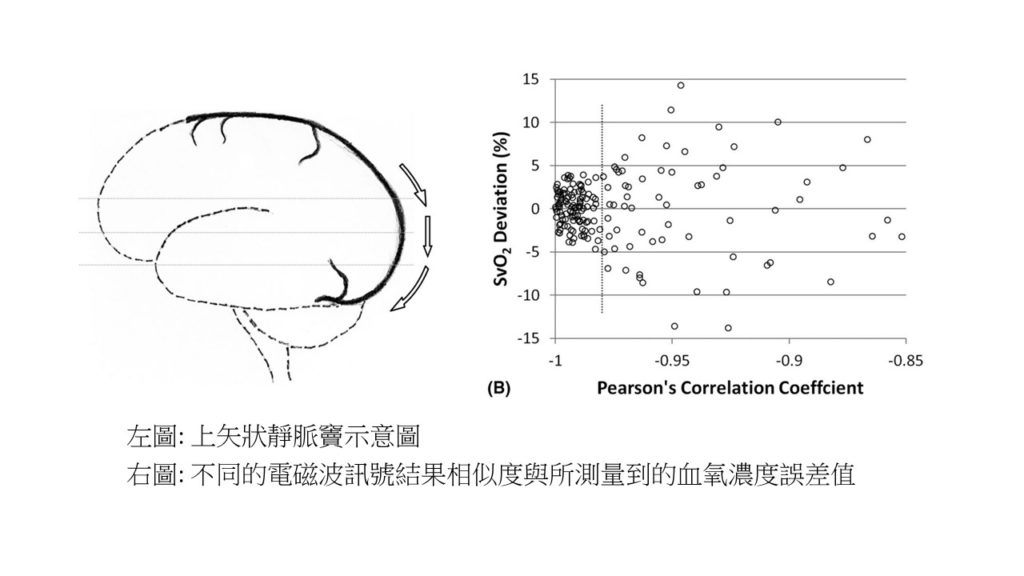
From the results of the article, when the similarity of the effects of different electromagnetic wave signals is more than 98%, the error value of the measured blood oxygen concentration does not exceed ! In other words, 3D multiple gradient-echo MRI can be used to measure SSS SvO2. Moreover, this method is advantageous in that it allows retrospective investigations on existing data whenever 3D multi-echo gradient-echo data have been acquired for the purpose of susceptibility-weighted angio/venography, QSM, or quantitative oxygenation venography. However, the SvO2 data could still be possibly contaminated by flow acceleration. Repeated acquisitions can only remedy this issue. In conclusion, using MRI to measure SSS SvO2 is still a significant breakthrough.
研究主題:醫學檢測技術:利用MRI測量大腦靜脈血氧濃度
撰寫:學生陳冠彬
大腦是一個很複雜的器官,他掌管著我們人體運作的所有功能,所以在醫療行為中檢測大腦的血氧濃度(SvO2)是相當重要的工程之一。在大腦的血管網路之中,負責將腦部深層的廢物帶出的血管稱作硬腦膜靜脈竇(Dural Venous Sinus),其中最大的靜脈即為上矢狀靜脈竇(Superior Sagittal Sinus, SSS)。而鍾老師的團隊利用了核磁共振搭配數學統計的方法來找出SSS中精準的血氧濃度數值。
根據鍾老師2020年五月發表在Magnetic Resonance in Medicine中的文章,利用3D多重梯度回聲法(3D multiple gradient-echo)來測量SvO2不僅是這種方法為非侵入式的手段,更是多數醫療團隊可以採用的方法,因為在磁敏感定量成像技術(Quantitative Susceptibility Mapping , QSM)中就經常使用這種方法。在文中亦有提到,利用3D多重梯度回聲法在測量時,最終影像的建構是透過回收不同的電磁波訊號再重新計算而成,但由於成像的過程中血液也同時在流動,所以需要在不同的收訊方向做流動補償(Flow compensation)才可以更完美的還原影像。
從研究結果指出,當不同的電磁波訊號結果相似度達98%以上時,所測量到的血氧濃度誤差值不超過!也就是說利用3D多重梯度回聲法來測量SSS中的SvO2是一可行的方法!另外,利用此分析技術更可以去重新回顧曾量測過得QSM影像中的SvO2。儘管如此,文中最後還是有談到此技術所面臨的問題,當掃描時遇到加速流動的狀況,還是會產生污染信號去影響最終的判讀,唯有不斷的反覆測量才有辦法補救。不過在現有技術之中,能夠利用MRI去實現造影跟量測已是重大的突破了。