研究主題-超音波導引注射輔具研發及深度學習於神經影像分割之應用
撰寫學生:陳冠彬
Point-of-care 指的是醫療人員隨時隨地都可以透過診斷工具來照顧病患,例如:血糖測試、懷孕檢測等。而隨著近年來Point-of-care 日益受到關注,超音波引導注射成為一項重要且受歡迎的技術。2019年郭教授在Journal of Clinical Medicine上發表了利用新型的二維雷射對準裝置(1.),以幫助臨床醫生在超音波引導注射中將針頭準確定位在掃描平面上。由於新手醫生在超音波引導注射的熟悉上往往需要大量的時間才可以掌握這項技術,例如,平面方法需要手眼協調訓練,以在掃描平面中準確定位針頭並以正確的角度推進,指向目標。所以開發能夠幫助新手更輕鬆的找到目標並改進他們的學習曲線的工具至關重要。目前市面上已有多種輔助對準裝置,但是仍舊有一些缺點,例如,Infiniti PlusTM針導系統一旦針頭插入組織就缺乏直接調整針頭角度的靈活性;電磁追踪系統、光學追踪系統和機器輔助系統太大且昂貴,無法在常規診所中使用。相反,基於雷射的輔助方法相對便宜且體積較小。然而,大多數最近開發的基於雷射的針導系統僅專注於協助在掃描平面中定位針頭,而未協助其插入角度。郭教授所開發的二維雷射對準裝置不但價格低廉、重量輕,易於製造,還可適應任何類型的超音波探頭。這項裝置透過不同模式的切換為針頭的定位提供引導,包括影像平面和插入的角度。這項研究大為縮短了新手掌握超聲波技術的時間,在超音波引導注射的培訓中提供了重大貢獻。
接著,今年5月,郭教授在Ultrasound in Medicine and Biology上發表了深度學習模型應用在神經超音波影像自動分割上的研究(2.)。腕隧道症候群( Carpal Tunnel Syndrome ,CTS )的患者,是因為在手腕水平處發生的正中神經( Median Nerve, MN )壓迫故造成病因,而動態超音波影像檢查在診斷以及治療CST患者上有著不可取代的重要性。郭教授的團隊成功利用深度學習模型在動態超音波影像中進行正中神經的影像分割。當患者在移動手指時,神經在橫截面上由於運動而變形和平移。被壓迫的神經會有多個形態特徵,如質心位移、橫截面面積和圓度。藉由檢測手指運動時受壓迫神經所呈現的異常特徵,可以幫助醫生判斷疾病嚴重程度和神經周圍注射的效果。但動態超音波影像的分析非常具有挑戰,在傳統方法中,要分析患者手指移動時的超音波影像需要仰賴大量人力來手動分割連續圖像。不僅如此,分割的準確性相當依賴檢查者的經驗水平,且在數據由不同經驗水平的檢查者處理或在高度變化的條件下獲取時,數據可能產生誤差,這也是為什麼深度學習的引入有其必要性。郭教授的團隊通過幾種最先進的端對端卷積神經網絡(CNN)模型對來自52名CTS患者的動態超聲檢查中的MN進行自動分割,並確定在基於單一幀進行訓練。最終成功利用此深度學習模型自動分割了動態超音波的影像,擺脫了以往需要大量人力才能完成的工作! 郭教授的團隊是領先使用深度學習模型在動態超音波影像中進行自動影像分割的團隊之一,也是第一個實現在動態超音波影像中對MN形態動態進行可視即時自動提取的團隊。
郭教授的研究在臨床環境中非常有價值,因為它允許臨床醫生在影像獲取的同時立即進行診斷和治療決策。深度學習模型可以自動分析肌骨結構,以協助疾病診斷和評估;而二維雷射對準裝置,可以幫助醫生更精準地進行下針治療。
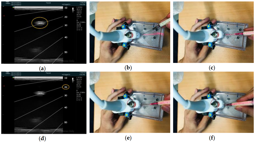
圖1. 二維雷射系統輔助超音波引導注射

圖2. 基於深度學習的動態超音波正中神經影像分割,不同顏色代表不同深度學習模型的預測結果,綠色為人工標註真實值
“Point-of-care” refers to the ability of healthcare professionals to provide patient care anytime and anywhere using diagnostic tools, such as blood glucose testing and pregnancy detection. In recent years, there has been a growing emphasis on point-of-care, and ultrasound-guided injection has emerged as an important and popular technique. In 2019, Professor Kuo published a study in the Journal of Clinical Medicine introducing a novel two-dimensional laser-align (2DLA) device (1.) designed to assist clinicians in accurately positioning the needle in the scan plane during ultrasound-guided injections. Novice physicians often require significant time to master ultrasound-guided injection techniques, particularly with in-plane approaches that demand hand-eye coordination for precise needle positioning and correct angulation toward the target. Therefore, the development of tools that facilitate novice access to the target is crucial to improving their learning curve.
Currently, various alignment devices are available in the market, but they often come with limitations. For example, the Infiniti PlusTM needle guidance system lacks flexibility for adjusting the needle angle once inserted into the tissue, and larger systems like electromagnetic tracking and optical tracking are impractical for regular clinics due to their size and cost. In contrast, laser-assisted approaches are relatively inexpensive and compact. However, many existing laser-based systems focus only on guiding needle positioning in the scan plane, neglecting assistance in insertion angle.
Professor Kuo’s 2DLA device addresses these limitations. It is affordable, lightweight, easy to manufacture, and compatible with any type of ultrasound transducer. The device offers guidance in three modes: 2DLA assisting, one-dimensional (1D) in-plane laser-align assisting without guidance for needle insertion angle, and no laser-align assisting, representing the traditional freehand approach. The research demonstrates that the 2DLA mode significantly reduces the time for successful targeting and retargeting frequency compared to the freehand (FH) and 1DLA modes. Rapid and accurate injection is a challenging aspect of ultrasound-guided injection training, and Professor Kuo’s device shows great potential in reducing the learning time required to master this skill and expediting the overall procedure.
Continuing this innovative work, Professor Kuo published a study in May of this year in Ultrasound in Medicine and Biology (2.), applying deep learning models to automatic segementation of nerve in dynamic ultrasonography. This research focused on patients with Carpal Tunnel Syndrome (CTS), where compression of the median nerve (MN) at the wrist level causes the condition. Dynamic ultrasound imaging is invaluable for diagnosing and treating CTS, providing real-time visualization of nerve deformation and translation during finger movement.
Professor Kuo’s team successfully employed deep learning models for the automated segmentation of the median nerve in dynamic ultrasound images. Abnormal features of the compressed nerve during finger movement, such as centroid displacement, cross-sectional area, and circularity, were detected to assist in evaluating disease severity and the effectiveness of perineural injections. Traditional manual segmentation of dynamic ultrasound images is labor-intensive and dependent on the examiner’s experience, leading to variability in data reproducibility. The introduction of deep learning addresses these challenges, offering an automated and consistent segmentation method.
The clinical significance of Professor Kuo’s research lies in its ability to enable immediate diagnosis and treatment decisions for clinicians during image acquisition. Deep learning models can automatically analyze musculoskeletal structures to aid in disease diagnosis and evaluation. Additionally, the 2DLA device enhances precision in needle treatments. Professor Kuo’s team is at the forefront of utilizing deep learning models for automated image segmentation in dynamic ultrasound, marking a significant advancement in real-time extraction of dynamic MN morphology.
In summary, Professor Kuo’s research contributes valuable tools and methodologies to the clinical environment. The combination of the 2DLA device and deep learning models holds promise for improving the efficiency and accuracy of ultrasound-guided procedures, ultimately benefiting both healthcare professionals and patients.
Ref:
- Shiao, E.C.; Kuo, P.-L. Two-Dimensional Laser-Align Device for Ultrasound-Guided Injection. J. Clin. Med. 2019, 8, 1048.
- Yeh, Cheng-Liang, et al. “Real-Time Automated Segmentation of Median Nerve in Dynamic Ultrasonography Using Deep Learning.” Ultrasound in Medicine & Biology 49.5 (2023): 1129-1136.